
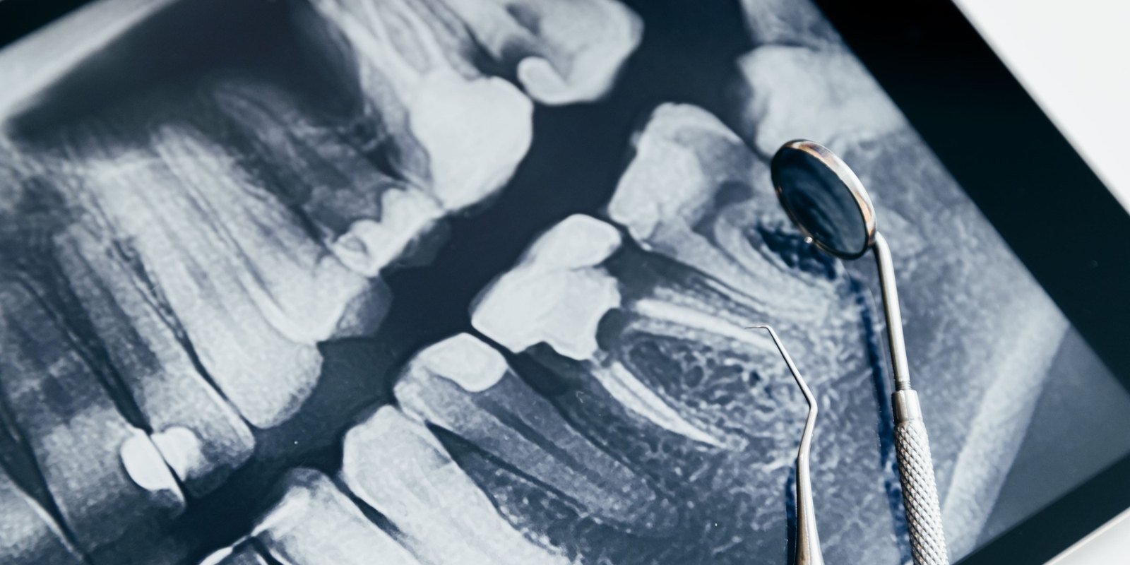
If you have ever been handed a lead apron and wondered whether you actually needed the images, the question is reasonable and the answer is not the same for everyone. Dental x-rays are not a routine box to tick at every visit.
This guide covers how often images are genuinely needed, what each type of x-ray is looking for, which personal factors shorten or lengthen the interval, and the specific practices that keep exposure low. It also covers the two situations patients ask about most: pregnancy and children.
There is no single schedule for dental x-rays. Adults with healthy teeth and no recent cavities may go two or three years between bitewings, while patients at higher risk of decay or gum disease need them more often. The interval is set by your history and current findings, not by the calendar.
How Often Dental X-Rays Are Actually Needed
There is no universal interval. The American Dental Association, in guidance developed with the U.S. Food and Drug Administration on selecting patients for dental radiographs, frames imaging as something ordered in response to clinical findings and patient history rather than on a fixed schedule. An adult with no recent decay, healthy gums and a stable history may go two to three years between routine bitewings. A patient with active decay may need them yearly or sooner, which is part of why the assessment at a preventive visit drives the imaging decision rather than the other way around.
New patients are a separate case. When a dentist has no prior records, a baseline set is usually taken so there is something to compare against later. That baseline is not repeated at the same frequency once it exists. If you have images from a previous practice, requesting them before your first appointment can avoid retaking what already exists.
The Main Types of Dental X-Ray and What Each One Shows
Different images answer different questions, which is why a dentist may take one type and not another. Knowing what each is looking for makes the recommendation easier to evaluate.
Bitewing x-rays
These show the crowns of the upper and lower back teeth in the same image, with the patient biting down on a tab. They are the workhorse for finding decay between teeth, where a visual exam and an explorer cannot reach, and for checking the bone level between the roots. Most routine recall imaging is bitewings.
Periapical x-rays
A periapical captures one or two teeth from crown to root tip, including the surrounding bone. This is the image taken when a specific tooth hurts, when an abscess is suspected, or when the dentist needs to see the root and the tissue at its tip. It is diagnostic rather than routine.
Panoramic x-rays
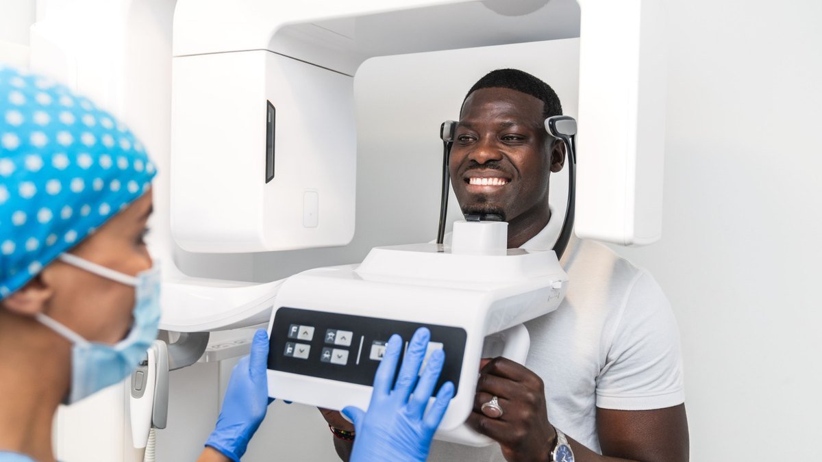
A panoramic image sweeps around the head and produces a single wide view of both jaws, the sinuses, the joints and any developing teeth. It trades fine detail for coverage, so it does not replace bitewings for spotting small cavities, but it is the image used for evaluating wisdom teeth, jaw injuries and broader structural questions. Imaging protocols, sterilization and the standards behind them sit under our technology and safety practices.
What Changes Your Recommended Interval
Risk is the variable that moves the schedule. A patient who has not had a cavity in a decade and one who has had three in two years are not on the same interval, and neither should be.
Factors that shorten the interval include a recent history of decay, existing large restorations, dry mouth from medication or medical conditions, a high sugar or high acid diet, smoking, orthodontic appliances that trap plaque, and a history of periodontal disease. That last one matters because bone loss is visible on imaging well before it is obvious any other way, which is why periodontal treatment relies on comparing images over time rather than a single snapshot.
Factors that lengthen it include a long stretch with no new decay, stable gum measurements, good home care and few existing restorations. Children and teenagers generally fall on the shorter end regardless, because their teeth are actively developing and enamel on newly erupted teeth is more vulnerable.
Symptoms override the schedule entirely. Pain, swelling, a broken tooth or an injury all call for imaging when they happen, not at the next scheduled visit.
How Dentists Keep Radiation Exposure Low
Dental radiography uses a small amount of ionizing radiation, and the exposure from a routine set is a small fraction of what a person receives from natural background sources over the course of a year. That does not make the amount irrelevant, which is why the field works from a principle known as ALARA, short for as low as reasonably achievable.
In practice ALARA means several things at once: taking only the images that will change a clinical decision, collimating the beam so it covers the area being imaged and no more, using protective shielding where it is appropriate, and holding the exposure settings to the minimum that still produces a readable image. It also means not repeating images that already exist and remain current.
Patients are entitled to ask why a specific image is being taken and what the dentist expects it to show. A clear answer is a reasonable thing to expect, and the dentist you see should be able to give it without hedging.
X-Rays During Pregnancy and for Children
Pregnancy is the most common reason patients ask to postpone imaging. Routine images that can wait are generally deferred, but dental care during pregnancy is not something to avoid, and an infection left untreated carries its own risks. When imaging is needed to diagnose a problem, protective shielding and limited exposure make it a considered decision rather than an automatic no. Tell your dentist you are pregnant or may be, before the appointment rather than in the chair.
For children, imaging tends to be more frequent than for low risk adults, because developing teeth change quickly and decay progresses faster through thinner enamel. Images also show whether permanent teeth are forming and erupting in the right position, which is often the first signal that orthodontic timing needs attention. For a child in pain or after a fall, imaging usually happens the same day as part of urgent dental care.
Schedule an exam with our Orlando team and we will review your history before recommending anything.
Frequently Asked Questions
Do I need dental x-rays every year?
Not necessarily. Yearly bitewings are common for patients at moderate or higher risk of decay, but adults with no recent cavities and stable gum health often go longer. The interval comes from your clinical history and current findings, so ask your dentist what puts you in the category they have assigned.
Can I refuse dental x-rays?
You can decline, and your dentist should explain what cannot be assessed without them. Decay between teeth, bone level changes and problems below the gumline are largely invisible on a visual exam alone. Declining is a choice to accept that limitation, so it is worth having the conversation rather than simply saying no.
Are dental x-rays safe?
The exposure from a routine dental set is small relative to the radiation people encounter from natural background sources in daily life. Dental offices work from the ALARA principle, taking only images that will change a decision and limiting exposure with shielding, collimation and minimum settings. No exposure is treated as trivial, which is why the imaging is selective.
How long are dental x-rays good for?
It depends on the type and your risk profile. Bitewings used for decay detection lose relevance as new decay develops, so a set from three years ago may not reflect your current situation. Panoramic images used for structural assessment often stay useful longer. Your dentist will tell you whether existing images still answer the question.
Can I bring x-rays from another dentist?
Yes, and it is worth doing. Requesting your records before a first appointment can prevent retaking images that already exist and gives the new dentist a comparison point, which is often more useful than a single current set on its own.
What can a dentist see on an x-ray that they cannot see in my mouth?
Decay between teeth and under existing fillings, bone loss from periodontal disease, abscesses at the root tip, cysts, impacted or extra teeth, and the position of teeth that have not erupted yet. Those are the findings that drive most treatment decisions, and nearly all of them are invisible during a visual exam.